Chest Muscle Anatomy Diagram - Amazon Com Male Chest Muscles Labeled Educational Medical Chart Black Wood Framed Art Poster 20x14 Home Kitchen / We think this is the most useful anatomy picture that.
Chest Muscle Anatomy Diagram - Amazon Com Male Chest Muscles Labeled Educational Medical Chart Black Wood Framed Art Poster 20x14 Home Kitchen / We think this is the most useful anatomy picture that.. 1300 x 1390 jpeg 297 кб. Human muscle system, the muscles of the human body that work the skeletal system, that are under voluntary control, and that are concerned with movement, posture, and balance. See more ideas about muscle diagram, medical anatomy, muscle anatomy. Human anatomy diagram shoulder anatomy shoulder muscles shoulder muscles and chest. Atlas of anatomy of the human body: Learn anatomy faster and remember everything you learn. Anatomical diagram showing a front view of muscles in the human body. When using a barbell they help explain the body's motions in really simple terms with diagrams. Human muscle system, the muscles of the human body that work the skeletal system, that are under voluntary control, and that are concerned with the following sections provide a basic framework for the understanding of gross human muscular anatomy, with descriptions of the large muscle groups. You may also find triceps, lateral head brachialis anatomynote.com found chest muscle anatomy from plenty of anatomical pictures on the internet.
The dominant muscle in the upper chest is the pectoralis major. Surrounding the rotator cuff muscles are many groups of muscles that work together to produce the various movements of the shoulder. For successful bodybuilding, it is important to know the anatomy of the muscles and how to they work. O muscles—sternocleidomastoid, anterior and middle scalene, infrahyoid, pectoralis major and minor, deltoid, trapezius, infraspinatus, supraspinatus, subscapularis diagrams of normal airway anatomy, lateral views. Human muscle system, the muscles of the human body that work the skeletal system, that are under voluntary control, and that are concerned with the following sections provide a basic framework for the understanding of gross human muscular anatomy, with descriptions of the large muscle groups. Female chest muscle anatomy diagram ~ diagram. #chest muscle anatomy and exercises #chest muscle chart #chest muscle diagram workout #muscle diagram of chest #muscle diagram of the chest.
View, isolate, and learn human anatomy structures with zygote body.
Freetrainers.com has a vast selection of exercises which are used throughout our workout plans. The movement that results from contraction is called the action of the muscle. Human muscle system, the muscles of the human body that work the skeletal system, that are under voluntary control, and that are concerned with movement, posture, and balance. Anatomical diagram showing a front view of muscles in the human body. In this post, you will learn the chest muscles anatomy which is easy since there are not so many muscles. Barbells are great for developing overall strength in your pressing muscles. You may also find triceps, lateral head brachialis anatomynote.com found chest muscle anatomy from plenty of anatomical pictures on the internet. We think this is the most useful anatomy picture that. Note that the middle lobe bronchi are relatively anterior (right. I wondered if i could request something. Download human muscle anatomy diagram vector art.
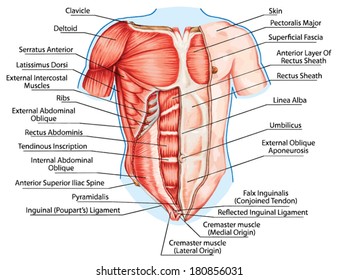
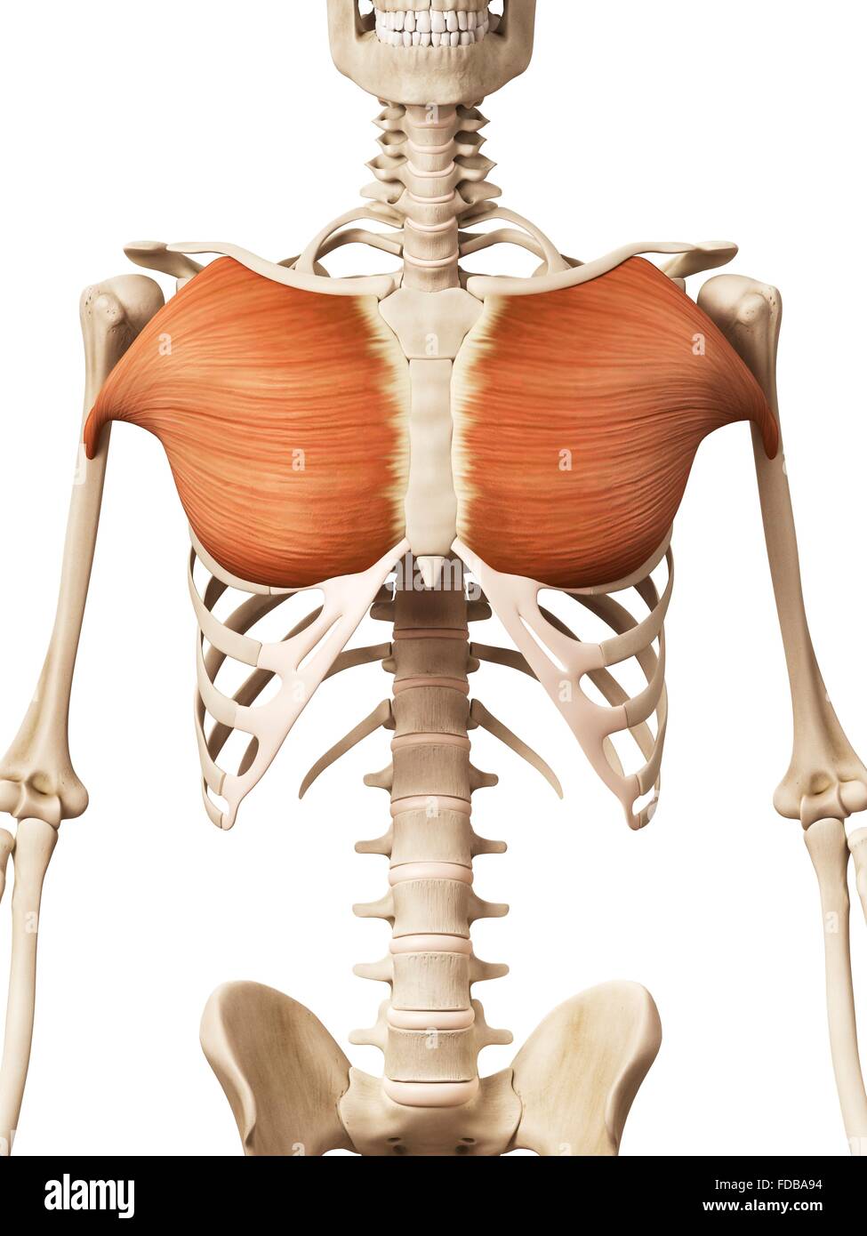
The chest can be split into two parts; See more ideas about muscle diagram, medical anatomy, muscle anatomy. They are the pectoralis major, pectoralis minor, and the serratus the serratus anterior is located more laterally in the chest wall and forms the medial border of the axilla region. You may also find triceps, lateral head brachialis anatomynote.com found chest muscle anatomy from plenty of anatomical pictures on the internet. To get started, choose a muscle group either on the muscle chart. Zygote body is a free online 3d anatomy atlas. The dominant muscle in the upper chest is the pectoralis major. Chest anatomy images, stock photos & vectors | shutterstock.
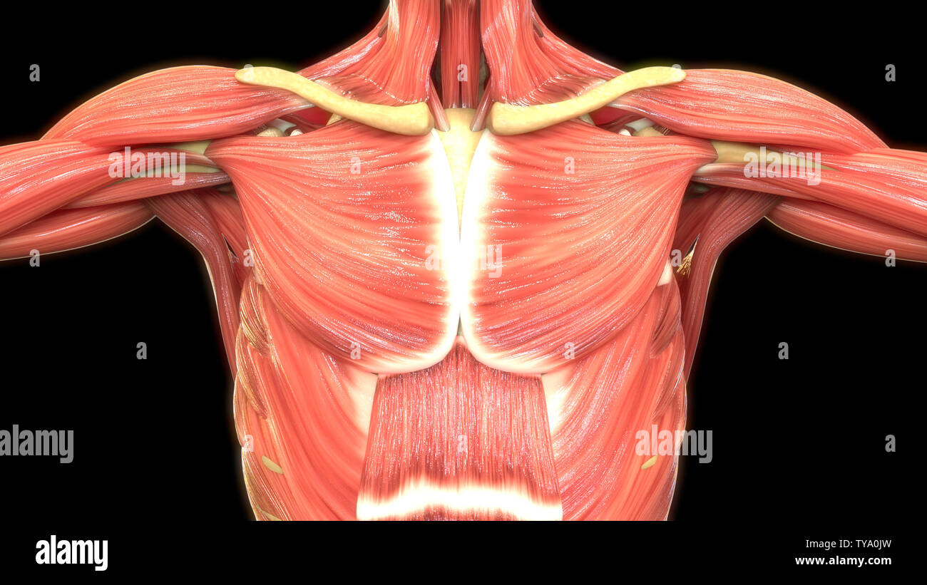
There are three muscles that lie in the pectoral region and exert a force on the upper limb.
O muscles—sternocleidomastoid, anterior and middle scalene, infrahyoid, pectoralis major and minor, deltoid, trapezius, infraspinatus, supraspinatus, subscapularis diagrams of normal airway anatomy, lateral views. Typically, one attachment remains stationary and is called the origin and the other attachment moves. Click on the labels below to find out more about your muscles. See more ideas about muscle diagram, medical anatomy, muscle anatomy. We think this is the most useful anatomy picture that. The chest anatomy includes the pectoralis major, pectoralis minor and the serratus anterior. Understanding the structure of a muscle fiber. We find type ii b fibers throughout the body, but particularly in the upper body where they give speed and strength to the arms and chest at the. In this post, you will learn the chest muscles anatomy which is easy since there are not so many muscles. 1300 x 1390 jpeg 297 кб.
It forms the bulk of the chest area and can be easily. O muscles—sternocleidomastoid, anterior and middle scalene, infrahyoid, pectoralis major and minor, deltoid, trapezius, infraspinatus, supraspinatus, subscapularis diagrams of normal airway anatomy, lateral views. I wondered if i could request something. When using a barbell they help explain the body's motions in really simple terms with diagrams. Freetrainers.com has a vast selection of exercises which are used throughout our workout plans. Anatomy • free medical books.
Learn anatomy faster and remember everything you learn.
So this is their bicep and it's flexing. I wondered if i could request something. 1300 x 1390 jpeg 297 кб. The two sides connect at the sternum, or breastbone. Chest anatomy images, stock photos & vectors | shutterstock. The chest can be split into two parts; Muscles that act on the chest. Download human muscle anatomy diagram vector art. Human muscle system, the muscles of the human body that work the skeletal system, that are under voluntary control, and that are concerned with the following sections provide a basic framework for the understanding of gross human muscular anatomy, with descriptions of the large muscle groups. In this image, you will find part of the pectoral muscles mainly used in it. Anatomical diagram showing a front view of muscles in the human body. Human anatomy diagram shoulder anatomy shoulder muscles shoulder muscles and chest. Learn anatomy faster and remember everything you learn.

Posting Komentar untuk "Chest Muscle Anatomy Diagram - Amazon Com Male Chest Muscles Labeled Educational Medical Chart Black Wood Framed Art Poster 20x14 Home Kitchen / We think this is the most useful anatomy picture that."